Posts

Showing posts from April, 2023

An Anatomy of Hand Fractures

Image
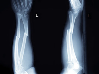
Several small bones together make up the supporting framework of the hand. These are the small finger bones, the phalanges, and the long bones, the metacarpals. Fracture in any of these bones is termed as a hand fracture. Various Type of Locking  orthopedic implants  are used for hand fracture surgery. More often Hand fractures are caused by an object either fall on the hand or the hand strikes an object occurs causing direct trauma to the hand. A twisting injury or a fall can also lead to a Hand fracture. Symptoms Some of the common indications of a hand fracture include: Pain Swelling A palpable deformity e.g. a shortened finger or a depressed knuckle Difficulty in moving the fingers A finger that crosses over the one next to it when one tries to make a fist. Diagnosis The doctor normally examines Hand for deformity, or problem in mobility, and lack of strength. A physical examination would be followed by x-ray to determine if a bone is broken. If a fracture is seen in one of the bon

Fracture Fixation Using Nails & Rods for Broken Bone

Image
Different types of orthopedic Implants have been designed for placement in the intramedullary canal of bones, ranging from Kirschner’s nails to the femoral nails. One can usually classify these devices by whether intramedullary reaming is essential prior to placement of the device or not. With the first nails placed down the femoral shaft, the medullary space first had to be reamed out so that the large nail wouldn’t shatter the bone as it is hammered down the shaft. However, reaming is an invasive process, and can compromise the already tenuous blood supply of the medullary space. Reaming can also lead to thermal osteonecrosis, especially if the medullary canal is small, a tourniquet is used while reaming, or there is marked soft tissue injury. If intraosseous pressure becomes elevated at the time of reaming, fat emboli to the lungs are possible. For these reasons, a different variety of un-reamed Implants have been developed. The Rush rod has a chisel-like tip, and is commonly used f

Uses of External Fixator Device for Bone Fracture

Image
External fixation is a method to provide stability to bone and soft tissue after a bone injury but can also be applied as a procedure to correct misalignment of bone, lengthen a limb, or protect soft tissue after a serious burn or injury. External fixation is carried out by fixing pins or screws into the bone on both sides of the fracture. The pins are clamped together outside of the skin and a series of clamps and rods form the external frame. It is generally done by an orthopedic surgeon under general anesthesia. Different steps of the procedure are: The bones around the fracture which are not damaged are drilled for fixing Pins/ Screws. Special threaded Pins are screwed into the holes. Outside of the body, rods with ball-and-socket joints are fixed with the bolts. The ball-and-socket joint can be adjusted to ensure the bone is aligned properly with as little, if any, shortening of a bone. The areas of skin pierced by the procedure must be cleaned regularly to prevent infection. In

Elbow Fracture Type – Cause, Symptoms & Treatment

Image
Elbow fractures mainly occur in the olecranon, the pointed bone at the elbow tip, where the bone cracks or breaks. This is a common injury that can be a part of a more severe condition or an isolated incident. If the bone breaks into parts and those parts become displaced within the elbow, this is called displaced fracture. A fracture in which bone sticks out by the skin is called an open fracture. The elbow joint comprises of bone, ligaments, cartilage, and fluid. Tendons and muscles help to move the elbow joint.  The three bones of elbow: the radius, the humerus (a bone in the upper arm) and the ulna (2 bones in the forearm). Symptoms Elbow fractures are often painful and constrict the range of movement. The area is often tender to the touch and moving it causes sudden, sharp pains. Other symptoms are: Instability in the joint, with the feeling that the elbow may “pop out” Swelling and bruising at the tip or back of the elbow Numbness in the fingers Bruising that travels down or up